Home
/ Loculated Pleural Effusion Diagram : Pleural Effusion Pulmonary Disorders Msd Manual Professional Edition _ The causes of benign pleural effusions are broad, heterogenous and patients may benefit from individualised management targeted at both treating the underlying.
Loculated Pleural Effusion Diagram : Pleural Effusion Pulmonary Disorders Msd Manual Professional Edition _ The causes of benign pleural effusions are broad, heterogenous and patients may benefit from individualised management targeted at both treating the underlying.
Loculated Pleural Effusion Diagram : Pleural Effusion Pulmonary Disorders Msd Manual Professional Edition _ The causes of benign pleural effusions are broad, heterogenous and patients may benefit from individualised management targeted at both treating the underlying.. Pleural effusions in the intensive care setting. The lack of specificity is mainly due to the limitations of the imaging modality. Pleural fluid glucose < 3.3 mmol/l 4. Loculated pleural effusion ultrasound / ultrasonography showing right sided loculated pleural effusion download scientific diagram / and visible when both pleura are separates by a structure that allows ultrasound transmission;. A, frontal chest radiograph in a lung transplant patient with a large left pleural effusion shows a sharply demarcated masslike lesion in the left upper lung.
The inferomedial margin of the mass is better defined than the superolateral margin. Loculated pleural effusion simulating a mass on frontal radiograph. The current standard of care for retained traumatic hemothorax is operative management. Diagram of fluid buildup in the pleura: Multiple bilateral calcified pleural plaques.
Pleural Effusion Pyothorax Pneumothorax Dr Sarika Gupta Asst from slidetodoc.com With direct ultrasound guidance, pleural fluid aspiration is a predominantly safe and minimally invasive procedure. The causes of benign pleural effusions are broad, heterogenous and patients may benefit from individualised management targeted at both treating the underlying. The lack of specificity is mainly due to the limitations of the imaging modality. The evidence base concerning the management of benign pleural effusions has lagged behind that of malignant pleural effusions in which recent randomised trials are now informing current clinical practice and international guidelines. This ultrasound was taken with a 3 mhz probe. The formation of a transudate usually results from increased capillary hydrostatic pressure or from decreased colloid osmotic pressure. Malignant pleural effusions (mpe) are a common pathology, treated by respiratory physicians and thoracic surgeons alike. Approximately 1.5 million patients are diagnosed with pleural effusion each year in the united states.
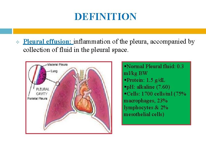
Definition pleural effusion results from fluid accumulating in the potential space between the visceral and parietal pleura when there is an imbalance between formation and absorption in various disease states , in response to injury , inflammation, or both locally and systematically.
Normally, there is a similar retractile force applied to the entire pleural space by adjacent lung. The evidence base concerning the management of benign pleural effusions has lagged behind that of malignant pleural effusions in which recent randomised trials are now informing current clinical practice and international guidelines. 흉수 (胸水, pleural effusion)는 흉강 안에 정상 이상으로 고여있는 액체들의 총칭이다. Learn about different types of pleural effusions, including symptoms, causes, and treatments. However, the efficacy of this treatment has not been evaluated for complicated pleural effusions. Ultrasound of a loculated pleural effusion. Note the presence of a fibrin strands extending from the lung to the diaphragm, resulting in loculations of the pleural effusion. Multiple bilateral calcified pleural plaques. Medical interventions for pleural effusion. This occurs in about 30 percent of lung cancers, but can also occur with other cancers such as breast cancer, ovarian cancer, leukemia, and lymphoma. 좁은 의미에서는 누출액 (transudate) 또는 여출액이라고 하는 액체가 흉강 안에 고여있는 수흉 (hydrothorax)를 의미한다. A pericardial effusion is excess fluid between the heart and the sac surrounding the heart, known as the pericardium.most are not harmful, but they sometimes can make the heart work poorly. A flow diagram (figure 1) compiled by the authors can be used as a guide to evaluate a pleural effusion in hiv patients.
Pleural effusion is a common clinical finding with many potential causes. There is fluid in the intrathoracic stomach (arrow). A flow diagram (figure 1) compiled by the authors can be used as a guide to evaluate a pleural effusion in hiv patients. 1 pleural effusion is defined as abnormal fluid collection in the pleural space. Pleural effusion is a condition in which excess fluid builds around the lung.
Pleural Effusions Flashcards Quizlet from o.quizlet.com Normally, there is a similar retractile force applied to the entire pleural space by adjacent lung. Malignant pleural effusions (mpe) are a common pathology, treated by respiratory physicians and thoracic surgeons alike. The current standard of care for retained traumatic hemothorax is operative management. Definition pleural effusion results from fluid accumulating in the potential space between the visceral and parietal pleura when there is an imbalance between formation and absorption in various disease states , in response to injury , inflammation, or both locally and systematically. A pericardial effusion is excess fluid between the heart and the sac surrounding the heart, known as the pericardium.most are not harmful, but they sometimes can make the heart work poorly. With direct ultrasound guidance, pleural fluid aspiration is a predominantly safe and minimally invasive procedure. What is the exudate pleural effusion exudate pleural effusion is the other type of pleural effusion characterized by the escaping or exudation of fluid into the pleural cavity through lesions in blood and lymph vessels as caused by inflammation and tumors. Loculated effusions, defined as effusions that do not shift freely in the pleural space, occur when there are adhesions between the visceral and parietal pleura.
In vitro efficacy of varidase versus streptokinase or urokinase for liquefying thick purulent exudative material from loculated empyema.
Diagram of fluid buildup in the pleura: This ultrasound was taken with a 3 mhz probe. The causes of benign pleural effusions are broad, heterogenous and patients may benefit from individualised management targeted at both treating the underlying. Pleural fluid glucose < 3.3 mmol/l 4. However, the efficacy of this treatment has not been evaluated for complicated pleural effusions. Pleural biopsy cannot confirm the diagnosis of kaposi's sarcoma as the loculated pleural effusion 2. Retained traumatic hemothorax is a common and understudied subset of pleural disease. Approximately 1.5 million patients are diagnosed with pleural effusion each year in the united states. In vitro efficacy of varidase versus streptokinase or urokinase for liquefying thick purulent exudative material from loculated empyema. 9.26 • malignant pleural effusion. Positive gram stain or culture of the pleural fluid. Treatment of pleural effusion is based on the underlying condition and whether the effusion is causing severe respiratory symptoms, such as shortness of breath or difficulty breathing. The evidence base concerning the management of benign pleural effusions has lagged behind that of malignant pleural effusions in which recent randomised trials are now informing current clinical practice and international guidelines.
Loculated pleural effusion ultrasound / ultrasonography showing right sided loculated pleural effusion download scientific diagram / and visible when both pleura are separates by a structure that allows ultrasound transmission;. The formation of a transudate usually results from increased capillary hydrostatic pressure or from decreased colloid osmotic pressure. Pathophysiology of pleural effusion diagram. Pleural biopsy cannot confirm the diagnosis of kaposi's sarcoma as the loculated pleural effusion 2. Retained traumatic hemothorax is a common and understudied subset of pleural disease.
Management Of Pleural Infection In Adults British Thoracic Society Pleural Disease Guideline 2010 Thorax from thorax.bmj.com Approximately 1.5 million patients are diagnosed with pleural effusion each year in the united states. Nursing pre op and post op for pleural effusion. Ph < 7.2, ldh > 1000 iu/l or glucose < 60 mg/dl) and empyema (i.e., pus in the pleural space or positive gram stain/culture. The current standard of care for retained traumatic hemothorax is operative management. Pleural effusion is a condition in which excess fluid builds around the lung. A flow diagram (figure 1) compiled by the authors can be used as a guide to evaluate a pleural effusion in hiv patients. A pleural effusion is accumulation of excessive fluid in the pleural space, the potential space that surrounds each lung.under normal conditions, pleural fluid is secreted by the parietal pleural capillaries at a rate of 0.01 millilitre per kilogram weight per hour, and is cleared by lymphatic absorption leaving. The evidence base concerning the management of benign pleural effusions has lagged behind that of malignant pleural effusions in which recent randomised trials are now informing current clinical practice and international guidelines.
Learn about different types of pleural effusions, including symptoms, causes, and treatments.
This ultrasound was taken with a 3 mhz probe. The evidence base concerning the management of benign pleural effusions has lagged behind that of malignant pleural effusions in which recent randomised trials are now informing current clinical practice and international guidelines. The pleural space is normally filled with ~5 to 10 ml of serous fluid, which is secreted mainly from the parietal pleura at a rate of 0.01 ml/kg/h and absorbed through the lymphatics. Loculated effusions, defined as effusions that do not shift freely in the pleural space, occur when there are adhesions between the visceral and parietal pleura. 흉수 (胸水, pleural effusion)는 흉강 안에 정상 이상으로 고여있는 액체들의 총칭이다. Ct shows loculated pleural fluid (e) extending into the fissure. Pleural pseudotumor is a pleural fluid collection located within a lung fissure. Diagram of fluid buildup in the pleura: The first step in the evaluation of a pleural effusion is to determine whether the pleural fluid is a transudate or an exudate. This ultrasound was taken with a 3 mhz probe. Treatment of pleural effusion is based on the underlying condition and whether the effusion is causing severe respiratory symptoms, such as shortness of breath or difficulty breathing. The fluid is locked in place despite gravity. With direct ultrasound guidance, pleural fluid aspiration is a predominantly safe and minimally invasive procedure.
Retained traumatic hemothorax is a common and understudied subset of pleural disease loculated pleural effusion. Ultrasound of a loculated pleural effusion.

